மனித உடற் செயன்முறைகள்
நீங்கள் சாதம் சாப்பிட்ட பின் அந்த உணவு வயிற்றில் மறைந்துபோவது ஏன்? நடக்கும்போது அதிகமாக மூச்சுவிடுவது ஏன்? வாழ்நாள் முழுவதும் ஓயாமல் இதயம் துடிப்பது எப்படி? கேள்வி கேட்டால் பதில் சொல்வது மூளையா, அல்லது உடல் முழுவதும் நீண்டிருக்கும் நரம்புகளா? நம் உடல் ஒரே நேரத்தில் ஐந்து பெரும் தொகுதிகளை ஒத்திசைவாகச் செயற்படுத்தும் மிகச் சிக்கலான இயந்திரம்: உணவுச் சமிபாட்டுத் தொகுதி, சுவாசத் தொகுதி, கழிவகற்றும் தொகுதி, குருதிச் சுற்றோட்டத் தொகுதி, இயைபாக்கமும் ஒர்சீர்த்திட நிலையமும் (homeostasis). இவ்வலகில் ஒவ்வொன்றினதும் அமைப்பு, தொழில், முக்கிய நோய்நிலைகள் ஆகியவற்றை ஆழமாக கற்போம்.
1. மனிதனின் உணவுச் சமிபாட்டுத் தொகுதி (Digestive system)
நாம் சாப்பிடும் உணவில் சாதம் (காபோவைதரேற்று), மீன், பயறு (புரதம்), நெய் (கொழுப்பு) ஆகியவை உள்ளன. செல்களுக்குப் பயனுள்ள எளிய மூலக்கூறுகளாக இவை மாற்றப்பட்டு உறிஞ்சப்பட வேண்டும். இது 2 படிகளில் நடைபெறுகிறது: (1) சமிபாடு (digestion), (2) உறிஞ்சல் (absorption). இவற்றை நிகழ்த்தும் உறுப்புகளின் கூட்டமே சமிபாட்டுத் தொகுதி (digestive system).
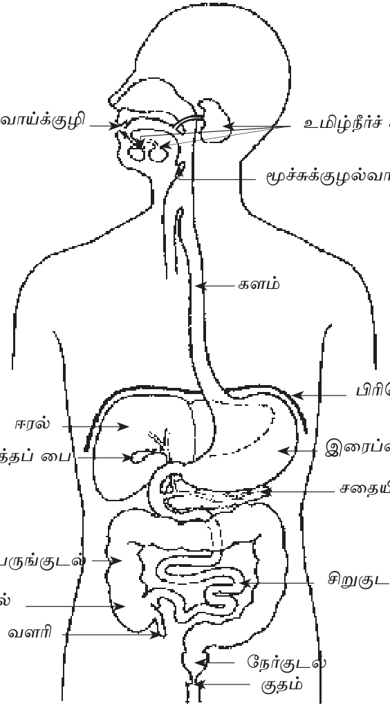
1.1 உணவுப் பாதையின் 6 பகுதிகள்
- வாய்க்குழி (Mouth): பற்களால் உணவு மெல்லப்படுகிறது (பொறிமுறைச் சமிபாடு); உமிழ்நீரிலுள்ள (saliva) ptyalin நொதி மாப்பொருளை (starch) மோல்ற்றோசாகச் (maltose) சிதைக்கத் தொடங்கும்.
- உணவுக்குழாய் (Oesophagus): குடலசைவு (peristalsis) மூலம் உணவு இரைப்பைக்குத் தள்ளப்படுகிறது.
- இரைப்பை (Stomach): HCl மற்றும் pepsin நொதி புரதத்தைப் பகுதியளவு சமிபாடடையச் செய்கின்றன. இங்குள்ள அமிலத்தன்மையால் நுண்ணுயிர்களும் கொல்லப்படுகின்றன.
- சிறுகுடல் (Small intestine): சதையீரல் சாறு, பித்தம், குடல் நொதிகள் ஆகியவை செயற்படுகின்றன. உறிஞ்சல் நிகழும் முக்கிய இடம் இதுவே; குடலுறிஞ்சிகளால் (villi) மேற்பரப்பளவு அதிகரிக்கிறது.
- பெருங்குடல் (Large intestine): நீரும் கனியுப்புகளும் உறிஞ்சப்படுகின்றன. இங்குள்ள பாக்டீரியாக்களால் வைட்டமின் K உம் சில B வகை வைட்டமின்களும் உற்பத்தியாகின்றன.
- நேர்குடல், குதம் (Rectum, anus): மலம் சேமிக்கப்பட்டு வெளியேற்றப்படுகிறது.
1.2 உதவி உறுப்புகள் (Accessory organs)
- ஈரல் (Liver): பித்தத்தை உற்பத்தி செய்கிறது (கொழுப்பைச் சிறு துளிகளாக்க உதவும்); குளுக்கோசைக் கிளைக்கோசனாகச் சேமிக்கிறது; நச்சுகளை நீக்குகிறது.
- பித்தப்பை (Gallbladder): பித்தத்தைச் சேமிக்கிறது.
- சதையீரல் (Pancreas): சமிபாட்டு நொதிகளையும் (amylase, lipase, trypsin) இன்சுலின் (insulin), குளுக்ககோன் (glucagon) ஹார்மோன்களையும் சுரக்கிறது.
- உமிழ்நீர்ச் சுரப்பிகள் (Salivary glands): உமிழ்நீரைச் சுரக்கின்றன.
1.3 மூன்று உணவு வகைகளும் அவற்றுக்கான நொதிகளும்
| உணவு | நொதி (Enzyme) | இறுதிப் பொருள் |
|---|---|---|
| Carbohydrate (starch) | Amylase, Maltase | Glucose |
| Protein | Pepsin, Trypsin | Amino acids |
| Fat (lipid) | Lipase | Fatty acids + Glycerol |
⭐ Quick rule Mouth = starch start; Stomach = protein start; Small intestine = all three complete + absorb.
2. சுவாசத் தொகுதி (Respiratory system)
Cells need O₂ to release energy from food (respiration: glucose + O₂ → CO₂ + H₂O + ATP). சுவாசத் தொகுதி O₂ ஐ உடலுக்குள் கொண்டுவருவதுடன், கழிவான CO₂ ஐயும் வெளியேற்றுகின்றது.
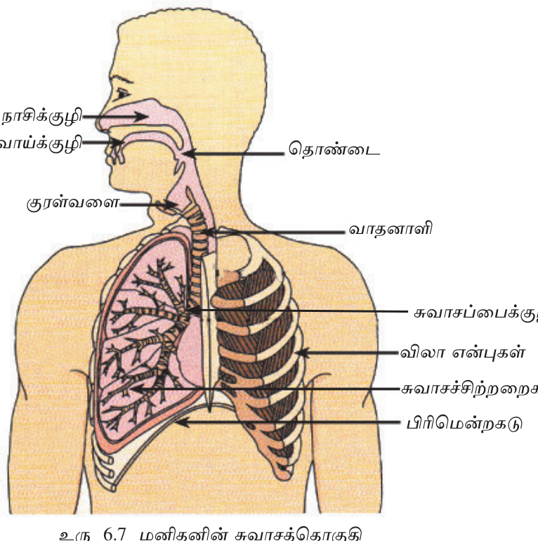
2.1 பாதை
நாசி → தொண்டை (pharynx) → குரல்வளை (larynx) → மூச்சுக்குழாய் (trachea) → மூச்சுக் கிளைக்குழாய்கள் (bronchi) → மூச்சுச் சிறுகுழாய்கள் (bronchioles) → நுரையீரல் (lungs) → நுண்காற்றறைகள் (alveoli; ஒரு நுரையீரலுக்கு ~300 million, வாயுப் பரிமாற்றம் நிகழும் சிறு பைகள்).
2.2 Alveoli — gas exchange site
நுண்காற்றறைகளின் (alveoli) சுவர் ஒரு கலம் தடிப்புடையது; அதைச் சுற்றி நுண்துளைக் குழாய்களின் (capillary) வலையமைப்பு உள்ளது. O₂ நுரையீரலிலிருந்து குருதிக்குள் விரவி, செங்குருதிக் கலங்களிலுள்ள ஈமோகுளோபினுடன் (haemoglobin) இணைகிறது; CO₂ எதிர்த்திசையில் விரவுகிறது. மொத்த நுண்காற்றறைப் பரப்பு ~70 m² ஆகும்; இது ஒரு டென்னிஸ் மைதானத்தின் பரப்புக்கு ஒப்பானது என்பது அதிசயமே!
2.3 Inhalation + Exhalation mechanism
- உள்ளிழுத்தல் (Inhalation): பிரிமென்றகடு (diaphragm) சுருங்கித் தட்டையாகிறது; விலாத் தசைகள் (intercostal muscles) விலா எலும்புகளை உயர்த்துகின்றன; இதனால் மார்புக் குழி விரிவடைகிறது. கனவளவு கூடுவதால் அழுத்தம் குறைந்து காற்று உள்ளே விரைகிறது.
- Exhalation: Reverse — diaphragm relax → dome up. Volume down → pressure up → air pushed out.
Normal adult: 12-20 breaths/min. Each breath ~500 mL. Daily volume ~10,000 L air.
3. கழிவகற்றும் தொகுதி (Excretory system)
வளர்சிதை மாற்றத்தால் (metabolism) கழிவுகள் (யூரியா, யூரிக் அமிலம், மிகையான உப்புகள், நீர்) உருவாகின்றன. கழிவகற்றும் தொகுதி, முக்கியமாக சிறுநீரகங்கள் (kidneys), இவற்றைக் குருதியிலிருந்து வடித்துச் சிறுநீராக வெளியேற்றுகின்றது.
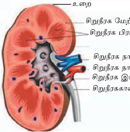
3.1 சிறுநீரகங்கள் (Kidneys): அவரை விதை வடிவிலான இணை உறுப்பு
- Each kidney has ~1 million nephrons (filtering units).
- Daily ~180 L blood filtered → ~1.5 L urine produced.
- Nephron 3 steps: glomerular filtration → tubular reabsorption → tubular secretion.
3.2 மற்றைய கழிவகற்றும் உறுப்புகள்
- Lungs: CO₂ + small water vapour.
- Skin: Sweat (water + salts + minor urea).
- Liver: Bile pigments — bilirubin (RBC breakdown).
4. குருதிச் சுற்றோட்டத் தொகுதி (Circulatory system)
இதயம் மையப் பம்பியாகவும், குருதிக் கலன்கள் விநியோக வலையமைப்பாகவும், குருதி கடத்தும் ஊடகமாகவும் செயற்படுகின்றன. இவை ஒட்சிசன், ஊட்டச்சத்துகள், ஹார்மோன்களைக் கலங்களுக்குக் கொண்டுசேர்த்து, CO₂ மற்றும் கழிவுகளை அகற்றுகின்றன.
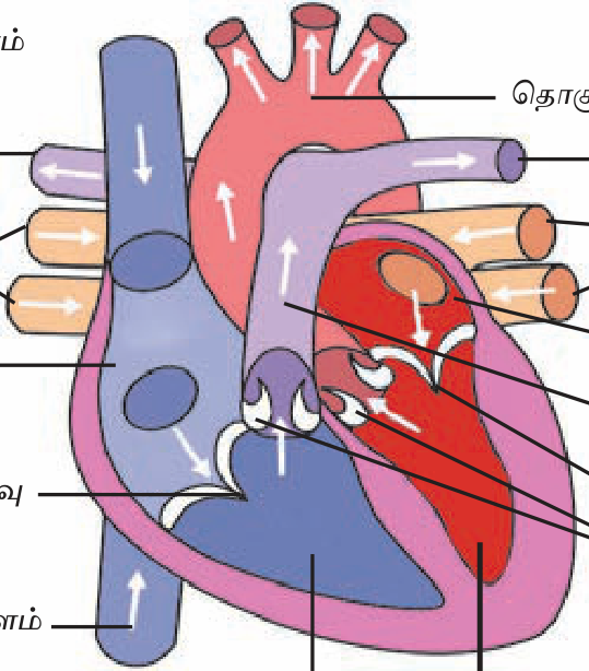
4.1 இதயம்: நான்கு அறைகளுடைய பம்பி
- வலது மேலறை (Right atrium): உடலிலிருந்து ஒட்சிசன் குறைந்த குருதியைப் பெறுகிறது.
- வலது கீழறை (Right ventricle): அதை நுரையீரலுக்கு அனுப்புகிறது (நுரையீரல் நாடி, pulmonary artery).
- இடது மேலறை (Left atrium): நுரையீரலிலிருந்து ஒட்சிசன் நிறைந்த குருதியைப் பெறுகிறது (நுரையீரல் நாளம், pulmonary vein).
- இடது கீழறை (Left ventricle): குருதியை உடல் முழுவதற்கும் அனுப்புகிறது (பெருநாடி, aorta); இதன் தசை மிகத் தடிப்பானது.
4.2 2 circuits
- Pulmonary (lungs): Right heart → lungs → left heart. Gas exchange.
- Systemic (body): Left heart → body → right heart. Nutrient + waste exchange.
4.3 Blood vessels
- Arteries: Thick walls, carry blood AWAY from heart (mostly oxygenated except pulmonary).
- Veins: Thin walls, valves, blood TO heart (mostly deoxygenated except pulmonary).
- Capillaries: One-cell thick, gas + nutrient exchange.
4.4 Blood components
- Plasma (55%): Liquid matrix, nutrients + hormones.
- RBC (45%): Haemoglobin → O₂ transport. ~5M/μL. No nucleus.
- WBC: Immunity. ~7K/μL. With nucleus.
- Platelets: Blood clotting.
Normal heart rate: 60-100 bpm at rest. Blood pressure: 120/80 mmHg.
5. இயைபாக்கமும் ஒர்சீர்த்திட நிலையமும் (Coordination and Homeostasis)
5.1 நரம்புத் தொகுதி (Nervous system)
மைய நரம்புத் தொகுதி (CNS) மூளையையும் முதுகுத்தண்டுவடத்தையும் கொண்டது; புற நரம்புத் தொகுதி (PNS) புற நரம்புகளைக் கொண்டது. இங்கு மின் சைகைகள் வேகமாகக் கடத்தப்படுகின்றன.
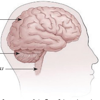
- பெருமூளை (Cerebrum): சிந்தித்தல், தன்னிச்சைச் செயல்கள், நினைவாற்றல்.
- சிறுமூளை (Cerebellum): சமநிலையைப் பேணுதல், ஒருங்கிணைப்பு.
- Brain stem: Heart, breathing, BP — involuntary controls.
- Spinal cord: Reflex arc + signal highway.
5.2 சுரப்பித் தொகுதி (Endocrine system)
Slower chemical signaling via hormones. Key glands:
- பிட்யூட்டரி (Pituitary, உச்ச சுரப்பி): "தலைமைச் சுரப்பி" (master gland). வளர்ச்சி ஹார்மோன் (growth hormone) உட்பட வேறு ஹார்மோன்களையும் சுரக்கும்.
- Thyroid: Metabolism, thyroxine. (Deficiency → goitre.)
- Pancreas: Insulin (lowers blood sugar) + glucagon (raises). Diabetes = insulin issue.
- Adrenal: Adrenaline (flight/fight).
- Gonads (ovary/testis): Sex hormones.
5.3 Homeostasis
Body temperature ~37°C; blood glucose ~5 mM; pH 7.4 — strict ranges maintained despite external variations. Negative feedback loops.
⚠ Disorders to know Diabetes (insulin failure), goitre (iodine deficiency → thyroid swelling), kidney failure (urea buildup), heart attack (coronary block), stroke (brain vessel block), hypertension.
✅ விரைவுச் சோதனை
முக்கியக் கருத்துக்களை உறுதிப்படுத்துங்கள். தவறான விடைகள் உங்கள் தவறுக் குறிப்பேட்டில் சேமிக்கப்படும்.
🖊 கட்டுரை வினாக்கள் (பகுதி II)
பரீட்சை வடிவில் கட்டமைப்பு வினாக்கள். முதலில் நீங்களே எழுதுங்கள்; பின்னர் மாதிரி விடையைத் திறந்து சரிபாருங்கள்.
விடைத் திட்டம் — சேர்க்க வேண்டிய புள்ளிகள்:
- 6 organs sequential: mouth, oesophagus, stomach, small intestine, large intestine, rectum.
- Liver — bile + storage + detox.
- Pancreas — enzymes + insulin.
- 3 macronutrient enzymes.
(1) வாய்க்குழி (Mouth): பல்லால் mechanical chewing → bolus formation. Salivary amylase (ptyalin) starch-ஐ maltose-ஆகச் சிதைக்கத் தொடங்கும். Tongue உணவை mix + swallow பயிற்சி.
(2) களம் (Oesophagus): 25 cm long muscular tube. Peristaltic contractions (wave-like) உணவை இரைப்பைக்கு push (gravity-independent — astronauts space-ல் சாப்பிட முடியும்).
(3) இரைப்பை (Stomach): J-shape muscular pouch. HCl + pepsin + gastric juice. pH ~1.5-2: (a) microbes kill, (b) pepsinogen → pepsin → protein partial digestion, (c) chyme உருவாக்கும். ~2-4 hour residence.
(4) சிறுகுடல் (Small intestine, 6 m): Three sections — duodenum (digestion), jejunum (absorption start), ileum (absorption continue). Bile (liver) + pancreatic juice + intestinal enzymes complete digestion. Villi + microvilli surface area ~200 m². Most nutrient absorption HERE.
(5) பெருங்குடல் (Large intestine, 1.5 m): Water + electrolyte absorption. Gut bacteria K + B vitamins synthesise. Faeces formation.
(6) நேர்குடல் + குதம் (Rectum + anus): Faeces store + voluntary elimination.
Accessory organs (உதவி உறுப்புகள்):
• ஈரல் (Liver, ~1.5 kg): Bile produces (fat emulsification). Glycogen storage. Drug + toxin detoxification. Plasma proteins (albumin, clotting factors) synthesise. Vitamin A, D, B12 storage.
• பித்தப்பை (Gallbladder): Liver-உற்பத்தி bile-ஐ store + concentrate; meal time-ல் duodenum-உக்கு release.
• சதையீரல் (Pancreas): Exocrine portion — amylase, lipase, trypsin enzymes secrete. Endocrine — insulin (lowers glucose) + glucagon (raises) hormones. Dual functions.
• உமிழ்நீர் சுரப்பிகள் (Salivary glands): 3 pairs — parotid, submandibular, sublingual. Daily 1-1.5 L saliva. Amylase + lysozyme + mucin.
3 macronutrient enzymes — final products:
• Carbohydrate (starch) → amylase + maltase → glucose.
• Protein → pepsin + trypsin + peptidase → amino acids.
• Fat → lipase (bile-emulsified) → fatty acids + glycerol.
All three absorbed at villi small intestine, transported via blood to liver (portal vein) for processing.
விடைத் திட்டம் — சேர்க்க வேண்டிய புள்ளிகள்:
- Air path: nose → pharynx → larynx → trachea → bronchi → bronchioles → alveoli.
- Inhalation mechanism.
- Alveolar surface 70 m², 1-cell wall, capillary network.
- O₂ in, CO₂ out by diffusion.
- Smoking damages ciliae + alveolar walls.
Air pathway:
நாசி (nostrils + nasal cavity) → pharynx (throat) → larynx (voice box) → trachea (windpipe) → 2 bronchi (one per lung) → progressively smaller bronchioles → terminal alveolar sacs.
Lungs: Spongy tissue, right lung 3 lobes, left lung 2 lobes (heart takes left space). Each lung ~300M alveoli. Total alveolar surface ~70 m² (tennis court size in chest cavity).
Breathing mechanism (inhalation):
• Diaphragm (dome muscle) contracts → flattens.
• External intercostal muscles raise ribs.
• Thoracic cavity volume ↑ → pressure ↓ (Boyle's law).
• Atmospheric air rushes in via mouth/nose.
Exhalation: Reverse — diaphragm relaxes, ribs lower, volume ↓, pressure ↑, air pushed out. Mostly passive (elastic recoil).
Normal rate: 12-20 breaths/min adult. Each ~500 mL (tidal volume). Total ~7 L/min at rest. During exercise → 100+ L/min.
Alveolar gas exchange:
Each alveolus = thin-walled sac (single cell thick) surrounded by dense capillary network (also single cell thick).
Diffusion (passive transport):
• O₂: Inhaled air ~21% O₂ → high partial pressure in alveolus. Capillary deoxygenated blood low pO₂. → O₂ diffuses alveolus → blood → binds Hb in RBCs.
• CO₂: Capillary blood high pCO₂ (cellular respiration waste) → low pCO₂ alveolus. CO₂ diffuses blood → alveolus → exhaled.
No energy needed — gradient-driven diffusion. ~300 mL O₂ absorbed/min at rest; ~250 mL CO₂ released.
Cellular use:
O₂ transported to all cells. In mitochondria: glucose + O₂ → CO₂ + H₂O + 36 ATP. ATP = cellular energy currency.
Smoking effects (புகைப்பழக்கம்):
• Cilia damage: Trachea + bronchi cilia (hair-like) mucus + debris sweep out. Smoke paralyses cilia → mucus accumulates → chronic cough.
• Alveolar destruction (emphysema): Inflammation breaks alveolar walls → surface area drops → less gas exchange → shortness of breath.
• Carcinogens: ~70 cancer-causing chemicals in tobacco smoke. Lung cancer 15-30× higher risk smokers.
• Tar deposition: Brown sticky residue darkens lungs.
• Carbon monoxide: CO binds Hb 200× stronger than O₂ → reduced O₂ delivery.
• Long-term: COPD, chronic bronchitis, heart disease, stroke, multiple cancers.
• Secondhand smoke harms others — children especially.
Sri Lanka tobacco prevention programmes since 2006 (NATA Act). Public smoking ban + warning labels.
விடைத் திட்டம் — சேர்க்க வேண்டிய புள்ளிகள்:
- 4 chambers: 2 atria + 2 ventricles.
- RA → RV → lungs (pulmonary).
- LA → LV → body (systemic).
- Valves prevent backflow.
- Systole + diastole.
- BP 120/80.
Human heart fist-sized muscular organ. Located in mediastinum (between lungs), slightly left.
Chambers:
• Right atrium (RA): Receives deoxygenated blood from body via superior + inferior vena cavae.
• Right ventricle (RV): Pumps deoxygenated blood to lungs via pulmonary artery. Thin muscular wall (low pulmonary resistance).
• Left atrium (LA): Receives oxygenated blood from lungs via 4 pulmonary veins.
• Left ventricle (LV): Pumps oxygenated blood to entire body via aorta. Thickest chamber (high systemic resistance).
Valves (4):
• Tricuspid: RA → RV. Prevents backflow.
• Pulmonary semilunar: RV → pulmonary artery.
• Mitral (bicuspid): LA → LV.
• Aortic semilunar: LV → aorta.
Valve failure → "murmur" sound. Rheumatic heart disease damages valves (post-streptococcal).
2 circulations:
(1) Pulmonary circulation (small):
RV → pulmonary artery → lungs → gas exchange → pulmonary veins → LA.
Deoxy → oxy in lungs.
Pulmonary artery = only artery carrying deoxygenated blood.
Pulmonary veins = only veins carrying oxygenated blood.
(2) Systemic circulation (large):
LV → aorta → systemic arteries → arterioles → capillaries (organ exchange) → venules → veins → vena cavae → RA.
Oxy → deoxy at tissues.
Cardiac cycle:
• Systole = ventricular contraction (pumping). ~0.3 s.
• Diastole = relaxation (filling). ~0.5 s.
Total cycle ~0.8 s → 75 bpm rest.
Blood pressure (BP):
• Systolic 120 mmHg = peak pressure during ventricular contraction.
• Diastolic 80 mmHg = minimum pressure during relaxation.
• Written: 120/80.
• Sphygmomanometer cuff + stethoscope measurement at brachial artery.
• Hypertension: >140/90 sustained = silent killer. Stroke, heart attack, kidney failure risk.
• Hypotension: <90/60 = fainting, shock risk.
ECG (Electrocardiogram):
Heart's electrical activity recorded. Waves:
• P wave: Atrial depolarization (contraction).
• QRS complex: Ventricular depolarization (powerful contraction).
• T wave: Ventricular repolarization (relaxation).
Normal ECG → heart electrical conduction healthy. Abnormalities:
• Arrhythmias (irregular rhythm)
• ST elevation → heart attack (myocardial infarction)
• Atrial fibrillation
• AV blocks
Life-saving diagnostic — emergency rooms standard.
விடைத் திட்டம் — சேர்க்க வேண்டிய புள்ளிகள்:
- 2 bean-shaped kidneys.
- 1M nephrons each.
- Filtration + reabsorption + secretion.
- 180 L filtered → 1.5 L urine.
- Failure → urea buildup → dialysis or transplant.
Two bean-shaped organs, ~12 cm each, retroperitoneal (behind peritoneum) at thoracolumbar level. Receive ~25% of cardiac output (~1.2 L/min blood) despite being only ~0.5% body weight.
3 layers:
• Cortex (மேற்படை): Outer layer, glomeruli located.
• Medulla (உள்படை): Inner pyramids, tubules.
• Renal pelvis: Collecting space draining into ureter.
Renal blood supply:
Abdominal aorta → renal artery → afferent arteriole → glomerulus → efferent arteriole → peritubular capillaries → renal vein → IVC.
Nephron — functional unit (~1 million per kidney):
(1) Bowman's capsule + glomerulus (filtration):
Glomerulus = ball of capillaries. High pressure → plasma pushed into Bowman's capsule. Cells + large proteins retained in blood; water + glucose + ions + small wastes (urea) pass through.
Filtration rate: ~125 mL/min = 180 L/day filtered.
(2) Tubular reabsorption (~99% recovery):
Filtrate flows through proximal convoluted tubule (PCT) → Loop of Henle → distal convoluted tubule (DCT) → collecting duct.
Useful substances actively/passively reabsorbed:
• Glucose 100% (PCT) — normally none in urine.
• Amino acids ~100%.
• Na+, K+, Cl-, HCO3- regulated.
• Water ~99% reabsorbed (controlled by ADH).
(3) Tubular secretion:
Blood → tubule: H+, K+, drugs, toxins actively secreted.
Final urine: ~1-2 L/day = 0.6% of filtered.
Urine composition (normal):
• 95% water
• Urea (main waste, ~2%)
• Salts (Na, K, Cl)
• Creatinine (muscle waste)
• NO glucose, NO proteins normally
Hormonal control:
• ADH (vasopressin): Pituitary, increases water reabsorption.
• Aldosterone: Adrenal, increases Na+ reabsorption.
• EPO (erythropoietin): Kidney-produced, stimulates RBC production.
Kidney failure (renal failure):
Causes:
• Diabetes (most common worldwide)
• Hypertension
• Chronic glomerulonephritis
• Polycystic kidney disease
• Drug toxicity (NSAIDs overuse, aminoglycosides)
• Sri Lanka specific: CKDu (Chronic Kidney Disease of unknown etiology) — agricultural communities particularly affected.
Consequences:
• Urea, creatinine, K+ buildup in blood (uraemia).
• Fluid overload → swelling, lung edema.
• Acidosis (H+ retention).
• Anaemia (no EPO).
• Bone disease (vitamin D activation failure).
Treatment options:
(1) Haemodialysis:
Artificial kidney machine. 3 sessions/week × 4 hr each typical. Blood pumped through dialyser (semipermeable membrane), waste diffuses into dialysate.
(2) Peritoneal dialysis:
Patient's peritoneal membrane used. Sterile fluid injected into abdomen, equilibrates with blood, then drained. Can be done at home.
(3) Kidney transplant:
Donor (living relative or deceased) kidney implanted. Best long-term outcome. Lifelong immunosuppression. Sri Lanka — National Hospital pioneers, NIROGI Lanka kidney foundation support.
Prevention:
Control diabetes + BP. Avoid NSAID overuse. Adequate hydration. Regular checkups (creatinine, urine analysis).
விடைத் திட்டம் — சேர்க்க வேண்டிய புள்ளிகள்:
- CNS = brain + spinal cord. PNS = nerves.
- Cerebrum, cerebellum, brain stem functions.
- Spinal cord reflex + signal.
- Neuron parts.
- Reflex arc: receptor → sensory → CNS → motor → effector.
- Pain withdrawal example.
2 divisions:
• CNS (Central Nervous System): மூளை + முதுகுத்தண்டுவடம் (Brain + spinal cord). Decision-making centre.
• PNS (Peripheral): Nerves connecting CNS to body. 12 cranial pairs + 31 spinal pairs.
Also functional divisions: Somatic (voluntary) + Autonomic (involuntary) → Sympathetic (fight/flight) + Parasympathetic (rest/digest).
Brain (3 main parts):
(1) Cerebrum (பெரு மூளை): 80% of brain mass. Folded surface (gyri + sulci) = extra area. 2 hemispheres (left + right) connected by corpus callosum.
4 lobes:
• Frontal: Planning, decision, personality, voluntary movement, speech (Broca's area).
• Parietal: Touch, temperature, pain sensation.
• Occipital: Vision processing.
• Temporal: Hearing, language understanding (Wernicke), memory (hippocampus).
(2) Cerebellum (சிறு மூளை): Below cerebrum, behind brain stem. Balance, coordination, fine motor skills, posture, motor learning. Damage = ataxia.
(3) Brain stem: Midbrain + pons + medulla. Autonomic vital functions — heart rate, breathing, BP, swallowing, vomiting reflexes. Damage often fatal.
Spinal cord:
Protected by vertebral column (33 vertebrae). 45 cm long. White matter (myelinated axons) outside, grey matter (cell bodies) inside (H-shape). Two functions:
• Carries signals up (sensory) + down (motor).
• Reflex centre (some don't need brain).
Neuron — functional unit:
• Cell body (soma): Nucleus + organelles.
• Dendrites: Receive signals from other neurons.
• Axon: Long extension, sends signal away. May be myelinated (faster) or unmyelinated.
• Synapse: Junction with next neuron. Neurotransmitter (acetylcholine, dopamine, etc.) chemical signaling.
3 functional types:
• Sensory (afferent): Receptor → CNS.
• Interneuron: Within CNS.
• Motor (efferent): CNS → effector (muscle/gland).
Reflex arc — uthaaranam:
Example: Pain withdrawal (சூடான பாத்திரம் தொட்டால்):
1. Stimulus: Hand touches hot vessel.
2. Receptor: Thermal/pain receptors in skin activated.
3. Sensory neuron: Carries signal via peripheral nerve to spinal cord.
4. Interneuron: Within spinal cord, processes + immediately relays to motor neuron (NO conscious brain processing).
5. Motor neuron: Sends signal back to arm muscles.
6. Effector: Biceps contracts → arm withdrawn.
7. Response: Hand pulled away — <0.1 seconds — even before pain felt!
Brain notification: Signal also ascends to brain via spinothalamic tract → conscious pain sensation + memory of "this is hot, avoid next time."
Why reflexes exist:
Speed = survival. Brain reaction ~0.3 s; reflex ~0.05 s. Protective.
Other reflex examples:
• Knee jerk (patellar): muscle spindle → spinal cord → quadriceps contract.
• Pupillary light reflex: bright light → pupil constrict.
• Blink reflex: corneal touch → eyelid closure.
• Salivation: food sight/smell.
Reflex testing: Doctors check spinal cord integrity. Absent/exaggerated reflexes signal neurological problems.
விடைத் திட்டம் — சேர்க்க வேண்டிய புள்ளிகள்:
- Pituitary, thyroid, pancreas, adrenal, gonads.
- Each hormone + function.
- Diabetes: insulin failure → high BG.
- Type 1 vs Type 2.
- Symptoms + complications + treatment.
Ductless glands secrete hormones directly into blood. Slower than nervous system but longer-lasting effects. Coordinates growth, metabolism, reproduction, stress response.
Major glands + key hormones:
(1) Pituitary (உச்ச சுரப்பி) — "Master gland":
Under brain (hypothalamus control). Pea-sized.
• Growth hormone (GH) → growth, metabolism.
• TSH → stimulates thyroid.
• ACTH → stimulates adrenal cortex.
• ADH → kidney water retention.
• Oxytocin → labour contractions + milk ejection.
• Prolactin → milk production.
• FSH, LH → gonad stimulation.
(2) Thyroid:
Butterfly-shaped, neck front. Requires iodine.
• Thyroxine (T4) + T3 → basal metabolic rate, body temp.
• Calcitonin → lowers blood calcium.
Disorders:
• Hypothyroidism → cold intolerance, weight gain, fatigue (Hashimoto's).
• Hyperthyroidism → weight loss, anxiety, tremor (Graves' disease).
• Goitre → iodine deficiency enlargement.
(3) Parathyroid:
Behind thyroid.
• PTH → raises blood calcium (bone, kidney).
(4) Pancreas:
Dual exocrine + endocrine. Islets of Langerhans:
• β cells → insulin (lowers BG).
• α cells → glucagon (raises BG).
(5) Adrenal:
On top of kidneys. 2 parts:
• Cortex → cortisol (stress, anti-inflammatory), aldosterone (Na/K balance), sex hormones.
• Medulla → adrenaline (fight/flight).
(6) Gonads:
• Testes → testosterone.
• Ovaries → oestrogen + progesterone.
(7) Pineal gland:
Melatonin → sleep-wake cycle (circadian rhythm).
(8) Thymus:
T-cell maturation (immunity).
----
Diabetes Mellitus (DM) — Detailed:
Definition: Chronic metabolic disorder of elevated blood glucose due to insulin deficiency, resistance, or both.
Normal glucose regulation:
After meal → BG rises → β cells secrete insulin → cells take up glucose → BG normalises. Liver stores excess as glycogen.
Between meals → BG falls → α cells secrete glucagon → liver releases glucose → BG maintained ~5 mM.
Diabetes types:
Type 1 (insulin-dependent):
• Autoimmune destruction of β cells.
• Usually onset childhood/young adulthood.
• Complete insulin deficiency.
• Always needs insulin injections.
• ~10% of all DM.
Type 2 (insulin-resistant):
• Cells resist insulin action + later β cell exhaustion.
• Usually onset adulthood (>40), but younger now due to obesity epidemic.
• Lifestyle-related — obesity, sedentary, diet.
• ~90% of all DM.
• Sri Lanka prevalence ~10% adults.
Gestational diabetes: Pregnancy-related. Often resolves after delivery but increases future risk.
Symptoms (3 Ps):
• Polyuria: Excessive urination (glucose pulls water).
• Polydipsia: Excessive thirst.
• Polyphagia: Excessive hunger (cells starved despite high BG).
• Plus: weight loss, fatigue, blurred vision, slow wound healing, recurrent infections.
Diagnosis:
• Fasting glucose ≥126 mg/dL (7 mM).
• Random glucose ≥200 mg/dL with symptoms.
• HbA1c ≥6.5% (3-month average).
• OGTT 2-hr ≥200 mg/dL.
Long-term complications ("Micro + Macro"):
Microvascular (small vessels):
• Retinopathy → blindness (leading cause worldwide).
• Nephropathy → kidney failure.
• Neuropathy → loss of sensation feet, autonomic dysfunction.
Macrovascular:
• Coronary artery disease → heart attack.
• Stroke.
• Peripheral arterial disease → amputation.
Other:
• Infections (poor immune response).
• Diabetic ketoacidosis (DKA) — emergency in Type 1.
• Cataracts.
Management:
(1) Lifestyle (Type 2 cornerstone):
• Diet: ↓ refined carbs, ↑ fiber, portion control.
• Exercise: 150 min/week moderate.
• Weight loss 5-10% body weight major improvement.
(2) Medications:
• Metformin (first-line Type 2).
• Sulphonylureas (insulin secretagogues).
• SGLT2 inhibitors, GLP-1 agonists (newer, also cardio-renal benefit).
• Insulin — all Type 1 + some Type 2.
(3) Monitoring:
• Self glucose monitoring.
• HbA1c every 3 months (target <7%).
• Annual: eye exam, foot exam, kidney function, lipids.
(4) Education + support:
• Diabetes educators.
• Family awareness.
• Sri Lanka — Diabetes Association support groups.
Prevention (Type 2):
Maintain healthy weight + regular exercise + balanced diet + avoid smoking. Family history → earlier screening recommended.
விடைத் திட்டம் — சேர்க்க வேண்டிய புள்ளிகள்:
- Definition.
- Negative feedback principle.
- Thermoregulation hypothalamus + sweat/shiver.
- Glucose: insulin/glucagon.
- Failure → hypothermia/hyperthermia, hypoglycaemia/diabetes.
The ability of organisms to maintain stable internal physical + chemical conditions despite external environmental variations. Coined by Cannon (1929) based on Bernard's "milieu intérieur" concept.
Examples of regulated variables:
• Body temperature: 37 ± 0.5 °C.
• Blood glucose: 4-7 mM.
• Blood pH: 7.35-7.45.
• Plasma osmolality: 280-295 mOsm/kg.
• Calcium: 2.2-2.6 mM.
• Oxygen saturation: 95-100%.
Negative feedback loop — core mechanism:
Components:
1. Stimulus: Change in regulated variable.
2. Sensor (receptor): Detects change.
3. Control centre: Integrates + compares to set point.
4. Effector: Acts to oppose the change.
5. Response: Variable returns to set point.
"Negative" = response opposes (reduces) the stimulus.
----
Example 1 — Thermoregulation:
Set point: ~37°C. Hypothalamus = control centre.
When body temp ↑ (hot day, exercise, fever):
• Sensor: Hypothalamic thermosensitive neurons + peripheral skin receptors detect ↑.
• Effectors:
- Sweat glands: Eccrine glands secrete sweat → evaporation cools (each gram water = 580 cal heat loss).
- Skin blood vessels: Vasodilation → more blood to surface → radiation heat loss.
- Behaviour: Seek shade, remove clothes.
• Result: Body temp ↓ back to 37°C.
When body temp ↓ (cold day):
• Sensor: Hypothalamic + skin cold receptors detect ↓.
• Effectors:
- Shivering: Skeletal muscle rapid contractions → heat production.
- Vasoconstriction: Skin blood vessels narrow → reduced heat loss.
- Goose bumps: Hair erector muscles (vestigial in humans).
- Brown fat thermogenesis: Babies (less developed in adults).
- Thyroid: Long-term ↑ metabolism.
- Behaviour: Wear more, seek warmth.
• Result: Body temp ↑ back to 37°C.
Fever:
Not homeostasis failure — controlled set-point elevation. Pyrogens (e.g., bacterial lipopolysaccharide → IL-1, IL-6) reset hypothalamus to higher set point. Body "feels cold" at 37°C, shivers to reach new set point ~39°C. Useful for fighting infection (most pathogens prefer 37°C).
Failures of thermoregulation:
• Hypothermia (<35°C): Confusion, slow breathing, eventually death. Cold exposure, immersion, alcohol intoxication.
• Hyperthermia (>40°C): Heat stroke. Brain damage, multi-organ failure, death if not rapidly cooled. Tropical climate + dehydration + exertion.
• Heat exhaustion: Excessive sweating → dehydration + electrolyte loss. Cramps, dizziness.
----
Example 2 — Blood glucose regulation:
Set point: 4-7 mM (~80-126 mg/dL fasting).
After eating (BG ↑):
• Sensor: Pancreatic β cells detect ↑ glucose.
• Effector: β cells secrete insulin.
• Insulin actions:
- Cells (muscle, fat) take up glucose via GLUT4 transporter.
- Liver stores glucose as glycogen.
- Inhibits gluconeogenesis.
- Promotes fat + protein storage.
• Result: BG ↓ back to set point.
Between meals (BG ↓):
• Sensor: Pancreatic α cells detect ↓ glucose.
• Effector: α cells secrete glucagon.
• Glucagon actions:
- Liver breaks down glycogen → glucose (glycogenolysis).
- Liver produces new glucose from amino acids (gluconeogenesis).
- Adipose tissue releases fatty acids.
• Result: BG ↑ back to set point.
Cortisol + adrenaline: Stress hormones also ↑ BG (longer-term + emergency).
Failures:
• Hypoglycaemia (<3.5 mM): Brain depends on glucose. Symptoms: sweating, tremor, confusion, seizure, coma. Insulin overdose, starvation, severe liver disease.
• Hyperglycaemia (chronic): Diabetes mellitus (covered separately). Long-term tissue damage.
• DKA: Severe insulin deficiency → fat breakdown → ketone bodies → metabolic acidosis. Emergency.
----
Other homeostasis examples (brief):
• pH: Buffer systems (bicarbonate) + lung CO₂ excretion + kidney H+ excretion.
• Water balance: ADH (vasopressin) + thirst.
• Calcium: PTH + calcitonin + vitamin D + kidney + bone.
• Blood pressure: Baroreceptors + heart rate + vascular tone + RAAS.
Why homeostasis matters:
Enzymes work optimally at narrow conditions. Outside range → cells die. Humans evolved set points for survival/reproduction. Disease often = homeostatic disruption.
🔥 மீட்டல் மையம்
பரீட்சைக்கு முன் இறுதி ஒரு நிமிடம் — மறக்கக்கூடாதவை மட்டும்.
- 5 body systems: Digestive · Respiratory · Excretory · Circulatory · Nervous/Endocrine (coordination).
- Digestion path: வாய் → களம் → இரைப்பை → சிறுகுடல் → பெருங்குடல் → குதம்.
- 3 macros: Carb → amylase → glucose. Protein → pepsin/trypsin → amino acid. Fat → lipase → fatty acid+glycerol.
- Accessory organs: ஈரல் (bile, store), பித்தப்பை (bile store), பாக்கிரியாஸ் (enzymes + insulin), salivary glands.
- Absorption: சிறுகுடல் villi-ல் primary.
- Respiration: நாசி → trachea → bronchi → bronchioles → alveoli (gas exchange).
- Inhalation: diaphragm ↓ contract, chest expand. Exhalation: reverse.
- Alveoli: 300M/lung, 70 m² surface, single-cell wall.
- Kidneys: 1M nephrons each, 180 L filtered → 1.5 L urine/day.
- Nephron 3 steps: filtration → reabsorption → secretion.
- Heart: 4 chambers (2 atria + 2 ventricles), 4 valves. BP 120/80.
- 2 circuits: Pulmonary (heart-lungs) + systemic (heart-body).
- Blood: Plasma 55% + RBC 45% + WBC + platelets.
- Brain: Cerebrum (thinking) + cerebellum (balance) + brain stem (vitals).
- Hormones: Insulin (pancreas, ↓BG), Adrenaline (stress), Thyroxine (metabolism), Growth hormone (pituitary).
- Homeostasis: Temp 37°C, BG 5 mM, pH 7.4 — negative feedback.
அலகின் முதுகெலும்பு — கருத்துக்களும் தொடர்புகளும்.
- 1. Digestive — mouth: Mastication + salivary amylase starch→maltose.
- 2. Stomach: HCl pH 2 (microbes kill) + pepsin protein digestion.
- 3. Small intestine (6m): Bile (liver, fat emulsify) + pancreatic enzymes + intestinal enzymes complete digestion. Villi+microvilli 200 m² absorption.
- 4. Large intestine: Water+mineral absorb. Bacterial vitamin K+B synthesis.
- 5. Liver: Bile, glycogen storage, detox, plasma proteins.
- 6. Pancreas: Exocrine (amylase, lipase, trypsin) + endocrine (insulin, glucagon).
- 7. Respiratory path: Nasal cavity → pharynx → larynx → trachea → bronchi → bronchioles → alveoli.
- 8. Breathing: Diaphragm contraction → volume↑ pressure↓ → inhale. Relaxation → exhale.
- 9. Gas exchange: O₂ alveolus→blood (Hb-bind in RBC). CO₂ blood→alveolus. Diffusion-driven.
- 10. Cellular respiration: Glucose + O₂ → CO₂ + H₂O + 36 ATP (mitochondria).
- 11. Kidney anatomy: Cortex + medulla + renal pelvis. ~1.2 L/min blood flow.
- 12. Nephron: Glomerular filtration (180 L/day) → tubular reabsorption (99%) → secretion. Final 1.5 L urine.
- 13. Urine: 95% water + urea + salts. NO glucose normally (Glycosuria = diabetes sign).
- 14. Other excretion: Lungs (CO₂), skin (sweat), liver (bile pigments).
- 15. Heart chambers: RA→RV→lungs (pulmonary); LA→LV→body (systemic). LV thickest.
- 16. Valves: Tricuspid, pulmonary, mitral, aortic. Prevent backflow.
- 17. Blood vessels: Arteries thick (high pressure) + Veins thin+valves (low return) + Capillaries 1-cell exchange.
- 18. Blood components: Plasma (yellowish liquid 92% water), RBC (Hb O₂ transport), WBC (immunity 5 types), Platelets (clotting).
- 19. Heart rate: 60-100 bpm rest. BP 120/80. Athletes 40-60.
- 20. CNS: Brain + spinal cord. PNS: peripheral nerves.
- 21. Cerebrum: 4 lobes (frontal-planning, parietal-touch, occipital-vision, temporal-hearing). Consciousness + memory.
- 22. Cerebellum: Balance, coordination, fine motor.
- 23. Brain stem: Vitals — heart, breathing, BP.
- 24. Spinal cord: Reflex centre + signal highway.
- 25. Neuron: Cell body + dendrites (input) + axon (output) + synapse. 3 types: sensory, inter, motor.
- 26. Reflex arc: Stimulus → receptor → sensory neuron → CNS interneuron → motor neuron → effector → response. <0.1 s.
- 27. Endocrine: Pituitary (master), thyroid (metabolism), pancreas (insulin), adrenal (stress), gonads (sex).
- 28. Diabetes: Insulin failure → high BG. Type 1 = β-cell destruction. Type 2 = insulin resistance.
- 29. Goitre: Iodine deficiency → thyroid enlargement.
- 30. Homeostasis: Negative feedback — stimulus→sensor→control→effector→opposite response.
பரீட்சைக்கு முந்தின இரவு முழு அலகையும் ஓட்டிப் பார்.
- Digestion: Mouth (amylase starch) → Stomach (HCl+pepsin protein) → Small intestine (complete + absorb) → Large intestine (water) → Rectum.
- Enzymes: Amylase=starch; Pepsin/trypsin=protein; Lipase=fat. End products glucose, amino acids, fatty acids.
- Accessory: Liver=bile+glycogen, Pancreas=enzymes+insulin, Gallbladder=bile store.
- Respiratory path: Nose→trachea→bronchi→bronchioles→alveoli (gas exchange).
- Inhale = diaphragm contracts ↓ + chest expands. 12-20 breaths/min, ~500 mL each.
- Alveoli: 300M/lung × 2, ~70 m² total surface, single-cell wall, capillary-rich.
- O₂ transport: RBC haemoglobin binds O₂ → tissues. CO₂ back to lungs.
- Cellular resp: C₆H₁₂O₆ + 6O₂ → 6CO₂ + 6H₂O + 36 ATP in mitochondria.
- Kidneys: Filter 180 L blood/day → 1.5 L urine. ~1M nephrons each.
- Nephron: Glomerular filtration → tubular reabsorption (99%) → secretion.
- Glucose in urine = diabetes. Protein in urine = kidney damage.
- Heart: 4 chambers (RA, RV, LA, LV). LV thickest = pumps body.
- RA→RV→lungs→LA→LV→body→RA. Pulmonary artery only artery with deoxy blood; pulmonary vein only vein with oxy.
- Vessels: Arteries thick away; veins thin+valves to heart; capillaries single-cell exchange.
- Blood: Plasma (55%) + RBC (O₂) + WBC (immune) + platelets (clot).
- BP normal: 120/80. Hypertension >140/90.
- Brain: Cerebrum (thinking, voluntary) + Cerebellum (balance) + Brain stem (heart, breathing).
- Spinal reflex: Pain → receptor → sensory → spinal interneuron → motor → muscle. Brain bypassed for speed.
- Hormones: Insulin lowers BG, Glucagon raises. Thyroxine ↑metabolism (iodine needed). Adrenaline = stress.
- Diabetes: ↑BG, polyuria, polydipsia, polyphagia. Long-term: retinopathy, nephropathy, neuropathy, CVD.
- Homeostasis: Temp 37°C, BG 5 mM, pH 7.4. Negative feedback maintains.
- ⚠ Common traps: Pancreas has BOTH digestion + hormones. Pulmonary artery carries deoxy blood. Reflex bypasses brain.
- ⭐ Numerical anchors: 4 chambers + 4 valves + 6 m small intestine + 300M alveoli + 70 m² alveolar surface + 180 L blood filtered + 1M nephrons + 5 L blood total + 5M RBC/μL.
- 📋 Tamil glossary: சமிபாடு=digestion; சுவாசம்=respiration; குருதி=blood; இதயம்=heart; சிறுநீரகம்=kidney; ஈரல்=liver; சதையீரல்=pancreas; நரம்பு=nerve; சுரப்பி=gland; ஹார்மோன்=hormone; மூளை=brain; ஒர்சீர்த்திட நிலையம்=homeostasis.